- 888 Normandy St.Houston, TX 77015
- + 1(832) 834-6678
- 6550 Fannin, Ste: 2403 Houston, Tx 77030
- + 1(713) 797-1111

CVVI - Cardiovascular & Vein Institute
Nuclear Stress Testing
Each year, more than 10 million nuclear stress tests are performed to diagnose and guide treatment in patients with heart disease. Nuclear stress tests use imaging to see how the heart performs both during physical activity and at rest, which can help diagnose heart disease, evaluate the severity of a heart condition, and help guide treatment plans. Although nuclear stress tests are generally safe, they do expose patients to radiation and slightly increase cancer risk in patients. Fortunately, there are many ways to help minimize exposure to radiation from nuclear stress tests, like by ensuring that the imaging is necessary and by using the safest imaging technique available. But according to a study published in the Journal of the American Medical Association, current practices may be exposing patients to unnecessarily high doses of radiation and increasing cancer risk.
More InformationCardiac Catheterization
Cardiac catheterization is a test to check your heart. This test can include a coronary angiogram, which checks the coronary arteries. A cardiac catheterization can check blood flow in the coronary arteries, check blood flow and blood pressure in the chambers of the heart, find out how well the heart valves work, and check for defects in the way the wall of the heart moves. In children, this test is used to check for heart problems that have been present since birth (congenital heart defect).
More InformationMagnetic Resonance Imaging
Magnetic resonance imaging (MRI) is a test that uses a magnetic field and pulses of radio wave energy to make pictures of organs and structures inside the body. In many cases MRI gives different information about structures in the body than can be seen with an X-ray, ultrasound, or computed tomography (CT) scan. MRI also may show problems that cannot be seen with other imaging methods.
More InformationCAT Scan
A conventional computerized axial tomography scan (CAT scan or CT scan) is an x-ray procedure which combines many x-ray images with the aid of a computer to generate cross-sectional views and, if needed, three-dimensional images of the internal organs and structures of the body. A CAT scan is used to define normal and abnormal structures in the body and/or assist in procedures by helping to accurately guide the placement of instruments or treatments.
More InformationChest X-ray
A chest X-ray is a radiology test that involves exposing the chest briefly to radiation to produce an image of the chest and the internal organs of the chest. An X-ray film is positioned against the body opposite the camera, which sends out a very small dose of a radiation beam. As the radiation penetrates the body, it is absorbed in varying amounts by different body tissues depending on the tissue's composition of air, water, blood, bone, or muscle. Bones, for example, absorb much of the X-ray radiation while lung tissue (which is filled with mostly air) absorbs very little, allowing most of the X-ray beam to pass through the lung.
More InformationCoronary Angiogram
An angiogram is an X-ray image of blood vessels after they are filled with a contrast material. An angiogram of the heart, a coronary angiogram, is the "gold standard" for the evaluation of coronary artery disease (CAD). A coronary angiogram can be used to identify the exact location and severity of CAD.
More InformationCT Scanning
Computerized (or computed) tomography, and often formerly referred to as computerized axial tomography (CAT) scan, is an X-ray procedure that combines many X-ray images with the aid of a computer to generate cross-sectional views and, if needed, three-dimensional images of the internal organs and structures of the body. Computerized tomography is more commonly known by its abbreviated names, CT scan or CAT scan. A CT scan is used to define normal and abnormal structures in the body and/or assist in procedures by helping to accurately guide the placement of instruments or treatments.
More InformationDoubutamine Stress Test
An echocardiogram (echo) is a test used to assess the heart's function and structures. A stress echocardiogram is a test done to assess how well the heart works under stress. The “stress” can be triggered by either exercise on a treadmill or medication called dobutamine. A dobutamine stress echocardiogram (DSE) may be used if you are unable to exercise. Dobutamine is put in a vein and causes the heart to beat faster. It mimics the effects of exercise on the heart.
More InformationECG
The electrocardiogram (ECG or EKG) is a noninvasive test that is used to reflect underlying heart conditions by measuring the electrical activity of the heart. By positioning leads (electrical sensing devices) on the body in standardized locations, information about many heart conditions can be learned by looking for characteristic patterns on the EKG.
More InformationECG Stress Testing
An exercise electrocardiogram (EKG or ECG) is a test that checks for changes in your heart while you exercise. Sometimes EKG abnormalities can be seen only during exercise or while symptoms are present. This test is sometimes called a "stress test" or a "treadmill test." During an exercise EKG, you may either walk on a motor-driven treadmill or pedal a stationary bicycle.
More InformationEchocardiographic Stress Testing (treadmill)
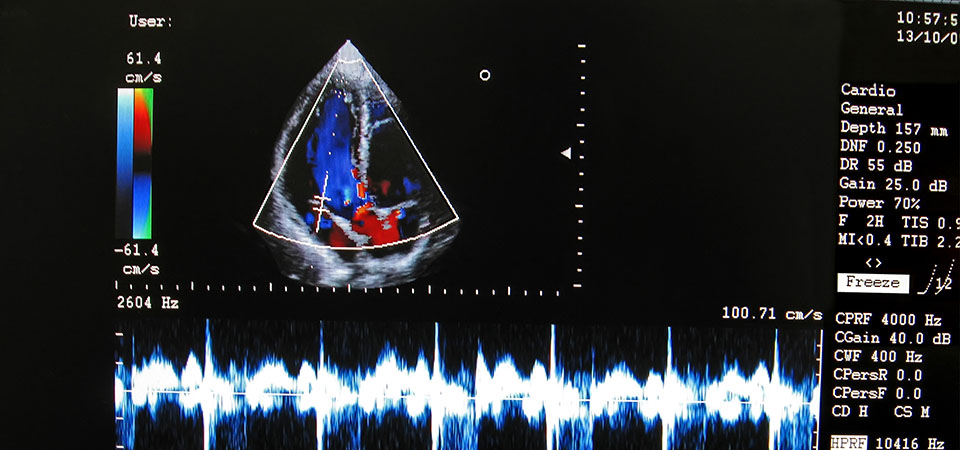
An echocardiogram is a painless, harmless test that uses high frequency sound waves (ultrasound) to examine the heart's anatomy and function.
More InformationEchocardiography
A supplement to the routine exercise cardiac stress test. During stress echocardiography, the sound waves of ultrasound are used to produce images of the heart at rest and at the peak of exercise.
More InformationHolter Monitor
In addition to the standard EKG, your doctor may recommend other specialized EKG tests, including a holter monitor or a signal-averaged electrocardiogram. A holter monitor is a portable EKG that monitors the electrical activity of a freely moving person's heart, generally for one to two days, 24 hours a day. It is most often used when the doctor suspects an abnormal heart rhythm or ischemia (not enough blood flow to the heart muscle).
More InformationMagnetic Resonance Imaging (MRI)
An MRI (or magnetic resonance imaging) scan is a radiology technique that uses magnetism, radio waves, and a computer to produce images of body structures. The MRI scanner is a tube surrounded by a giant circular magnet. The patient is placed on a moveable bed that is inserted into the magnet. The magnet creates a strong magnetic field that aligns the protons of hydrogen atoms, which are then exposed to a beam of radio waves. This spins the various protons of the body, and they produce a faint signal that is detected by the receiver portion of the MRI scanner. The receiver information is processed by a computer, and an image is produced.
More InformationMultiple Gated Acquisition (MUGA) Study
A multigated acquisition scan (also called equilibrium radionuclide angiogram or blood pool scan) is a noninvasive diagnostic test used to evaluate the pumping function of the ventricles (lower chambers of the heart). During the test, a small amount of radioactive tracer is injected into a vein. A special camera, called a gamma camera, detects the radiation released by the tracer to produce computer-generated movie images of the beating heart. The MUGA scan is a highly accurate test used to determine the heart’s pumping function.
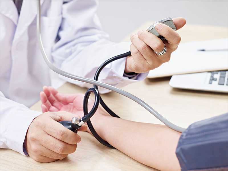
More InformationPhysical Exams
The physical exam is an essential part of any doctor's visit. Surprisingly, though, there are no absolutes in a routine physical. A good doctor may be thorough or brief, but he or she will spend time listening to your concerns and providing counseling for your particular needs.
More Information


- Nuclear Stress Testing
- Cardiac Catheterization
- Magnetic Resonance Imaging
- CAT Scan
- Chest X-ray
- Coronary Angiogram
- CT Scanning
- Doubutamine Stress Test
- ECG
- ECG Stress Testing
- Echocardiographic Stress Teting (treadmill)
- Echocardiography
- Holter Monitor
- Magnetic Resonance Imaging (MRI)
- Multiple Gated Acquisition (MUGA) Study
- Physical Exams

